


Bovine Besnoitiosis
Bovine besnoitiosis is a skin disease of cattle caused by the protozoan (single-celled) intracellular parasite Besnoitia besnoiti.
Bovine besnoitiosis is a notifiable disease in Ireland, where it has been detected in a small number of herds in recent years. There are no known food safety or human health risks associated with the disease. Besnoitiosis has been diagnosed over many years in many parts of the world and has never been reported to infect humans.
Bovine besnoitiosis is usually a mild low-impact disease, resolving over the course of 1-2 weeks. Affected cattle do not usually die of the condition although severely affected animals may have a prolonged recovery. Clinical signs appear during the summer and affected animals remain carriers for life. There are no effective drugs or vaccines currently available in Europe. Therapeutic intervention is largely limited to generic topical treatment of skin lesions, and vector (fly) control. Equally, there are few control measures that can be recommended other than fly control and maintaining a closed herd. Neither of these control measures is viewed as completely or 100% effective, because the disease can be carried by vectors including flies.
Bovine besnoitiosis has, for many years, been observed in Africa, the Middle East and more recently in Europe, where it has been seen since the 199’s – with cases reported in Spain, Portugal, France, Italy, Germany Switzerland, Hungary, Croatia, Greece, and Belgium. The full life cycle of this parasite has not been fully established and may involve wildlife, but cattle are the only domestic species that have been reported to show clinical disease. Mechanical transmission between cattle is believed to occur via insect vectors, with close contact between infected and healthy animals also considered quite important for disease transmission; and transmission directly between cattle via the naso-pharyngeal route has also been demonstrated. In addition, the use of hypodermic syringes for multiple animals will mechanically transfer infection between animals. There are no reports of vertical transmission from parents to offspring.
The disease displays a variety of presentations and infected cattle often show little or no obvious clinical signs; the initial acute phase clinical presentation occurs when the parasite (in the immature tachyzoite form) is rapidly multiplying and causing tissue damage particularly in the blood vessels of the dermis, subcutis, fascia, testes and upper respiratory mucosa. This stage usually presents as one or more of the following: fever, anorexia, rhinitis, weakness, milk drop, stiffness, abortion and thus many of the sick animals may superficially present a syndrome suggestive of a clinical case of IBR. The more chronic stage starts 1-2 weeks after the onset of the acute stage and clinical signs include intermittent fever, anorexia and weight loss. Most animals go on to show skin and eye lesions, which include thickening, hardening and folding or wrinkling of the skin especially around the neck, shoulders and rump. The udder may also be affected. There may be some swelling of the legs and animals may appear ‘stiff’ when moving. Laminitis is a complication in the minority of cases that progress to severe chronic disease. Small cysts are often visible in the sclera and conjunctiva of the eye, and vulva and these are very useful in clinical diagnosis. Some cases may superficially resemble photosensitisation.
The experience with besnoitiosis in this country and in other countries indicates that it is a relatively mild and slow-spreading disease, with exposed animals developing good immunity, not all in-contact animals becoming affected, most affected animals having minor lesions, with some young animals becoming infected and showing clinical disease as they join the herd each year.
Samples from clinical suspects for testing may be submitted to the Regional Veterinary Laboratories at Athlone, Backweston, Cork, Kilkenny, Limerick or Sligo. Testing is based on clotted blood samples for serological testing for antibodies (which confirms exposure) and /or histopathology of skin biopsies of suspect lesions (which confirms if an animal has the disease).
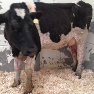
Picture 1: cow showing clinical signs of Besnoitiosis
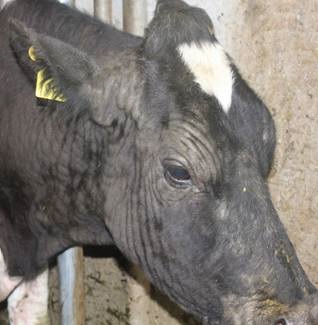
Image 2: cow showing clinical signs of Besnoitiosis
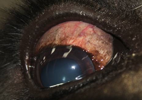
Image 3: eye lesion caused by Besnoitiosis
Images with thanks to the Regional Veterinary Labs.